Platecarpus tympaniticus is a Plioplatecarpinae Mosasaurid from late Cretaceous USA and Belgium. It was closely related to Plioplatecarpus and Igdamanosaurus.
Description[]
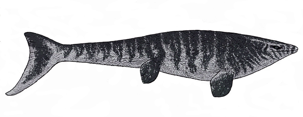
Platecarpus had a long, down-turned tail with a large dorsal lobe on it, steering flippers, and jaws lined with conical teeth. It grew up to 4.3 m (14 ft) long, with half of that length taken up by its tail. The platecarpine mosasaurs had evolved into the very specialized plioplatecarpine group by the end of the Cretaceous.
The skull structure of Platecarpus is unique among mosasaurs. This genus is characterized by a short skull, and has fewer teeth than any other mosasaur (around 10 teeth in each dentary).[note 1] LACM 128319 preserves matter within the sclerotic ring that may possibly be the retina of the eye. Small structures in the retina, each around 2 µm long and observed by scanning electron microspectroscopy, may represent retinal melanosomes preserved in their original positions.
Size comparison[]
The respiratory tube is also known in LACM 128319, preserved as cartilaginous tracheal rings. Only the posterior-most end of the tracheal tube – at the end of the neck near the pectoral girdle – is known. The section where the two bronchi split was also preserved in the specimen, but was destroyed during excavation. This is an indication that Platecarpus and other mosasaurs had two functional lungs. Snakes, which are closely related to mosasaurs, have only one functional lung with the second often being vestigial or absent. Unlike terrestrial lizards, however, the bronchi separate in front of the area of the forelimbs rather than at the level of the limbs.
Skin impressions are known from Platecarpus, preserved in LACM 128319 as soft impressions and phosphate material. Scales on the tip of the snout and the top of the skull are somewhat hexagonal in shape and do not touch one another. The scales on the jaws are longer and rhomboidal in shape, overlapping one another. The scales on the snout indicate that the nostrils were placed far in front of the skull at its tip and faced laterally as in most squamates and archosaurs. The body scales are all rhomboidal in shape and form tightly connecting diagonal rows that overlap each other at their posterior edges. They are generally the same size throughout the entire length of the body. The caudal scales on the tail are taller and larger than those of the rest of the body, although those covering the lower surface of the tail are more similar to body scales. Internal organs, or viscera, may also be preserved in the specimen as reddish areas. One is located in the thoracic cavity low in the ribcage, while the other is located in the upper portion of the abdominal cavity just behind the ribcage. The reddish areas were analysed with mass spectrometry and were shown to contain high levels of compounds made of iron and porphyrin. These substances are evidence of hemoglobin decomposition products that may have formed in the organs as they decomposed. Based on its position, the organ in the thoracic cavity is probably the heart or liver, or even both of those organs. The organ in the abdominal cavity may be a kidney, although it is in a more anterior position than the kidneys of monitor lizards, mosasaurs' closest living relatives. The anterior position of the kidneys may have been an adaptation toward a more streamlined body, as their presumed position is similar to that of cetaceans.
History[]
Various skeletons of this mosasaur have been found in Cretaceous deposits in Kansas, but only one complete skull has ever been recovered.[3] Platecarpus fossils have been found in rocks that date back to the late Coniacian through the early Campanian in the Smoky Hill Chalk.
Paleobiology[]
Compared to the tylosaurs, plioplatecarpine mosasaurs had much less robust teeth, suggesting that they fed on smaller (or softer) prey such as small fish and squid.
While mosasaurs are traditionally thought to have propelled themselves through the water by lateral ungulation in a similar way to eels, the deep caudal fin of Platecarpus suggests that it swam more like a shark. The downturned caudal vertebrae of Platecarpus suggest it had a crescent-shaped tail fluke. At the point of the tail where the fluke begins the vertebral centra are shortened and disk-like. Their reduced size likely allowed for greater flexibility at an area that would have experienced high stresses during swimming. The neural spines of these vertebrae also have grooves for the insertion of interspinal ligaments and dorsal connective tissues which would have aided in lateral movement of the fluke. The ligaments were probably made of collagenous fibers that acted as springs to move the tail back into a resting position after energy was stored in them. These types of ligaments work in some living fish to conserve energy during repetitive bending of the tail. While the fluke and back of the tail undulated in Platecarpus, the base of the tail remained stable. This form of movement is known as carangiform locomotion
The structure of the scales of Platecarpus may have been another adaptation toward a marine lifestyle. The small size and similar shape of these scales throughout the body would have stiffened the trunk, making it more resistant to lateral movement. This stiffness would have improved hydrodynamic efficiency by improving the flow of water across the body. The early mosasauroid Vallecillosaurus also preserves body scales, but they are larger and more varied in shape, suggesting that the animal relied on undulatory movement in its trunk rather than just its tail. Plotosaurus, a more derived mosasaur than Platecarpus, has even smaller scales covering its body, indicating that it had even more efficient locomotion in the water.
In popular culture[]
- Platecarpus appeared in the video game Jurassic Park Builder.
Gallery[]
Platecarpus/Gallery